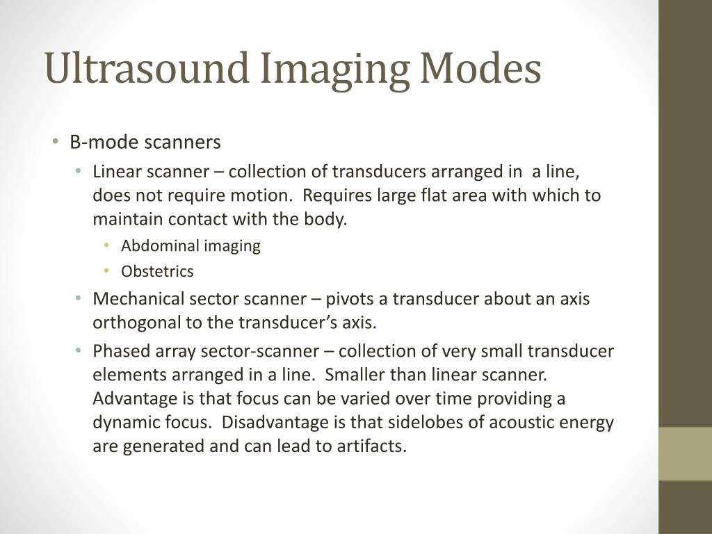
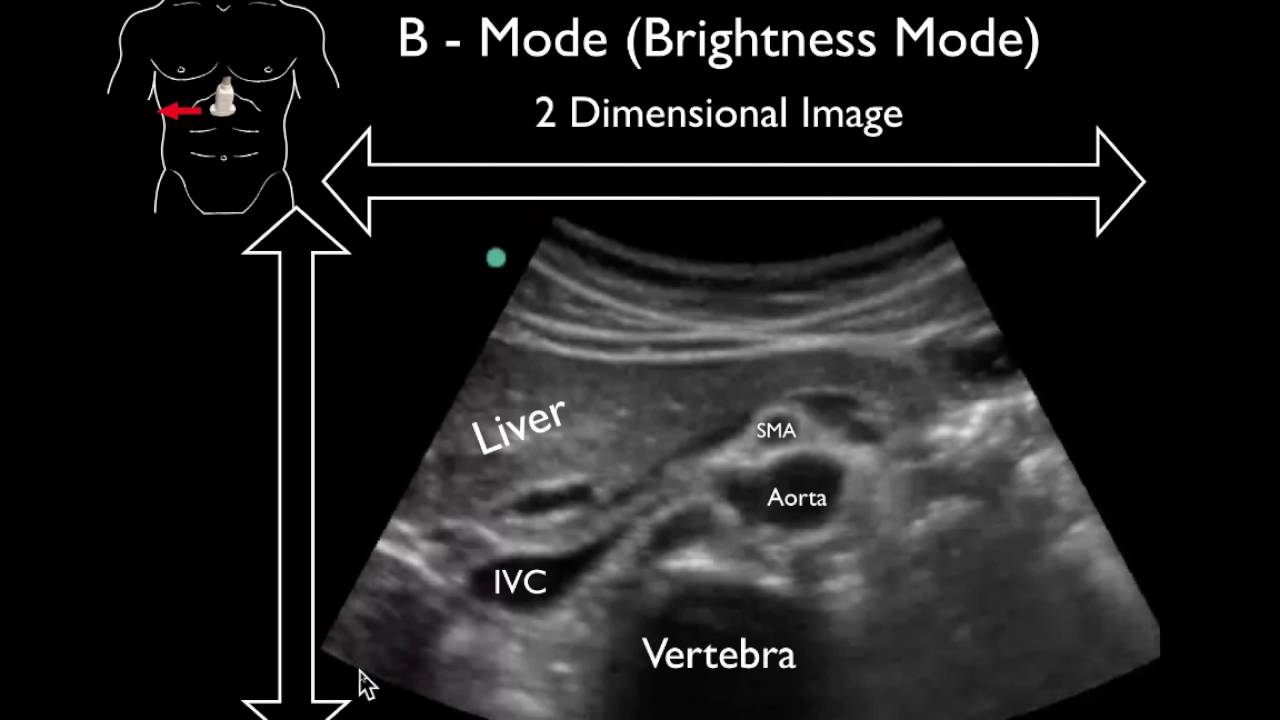
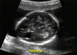
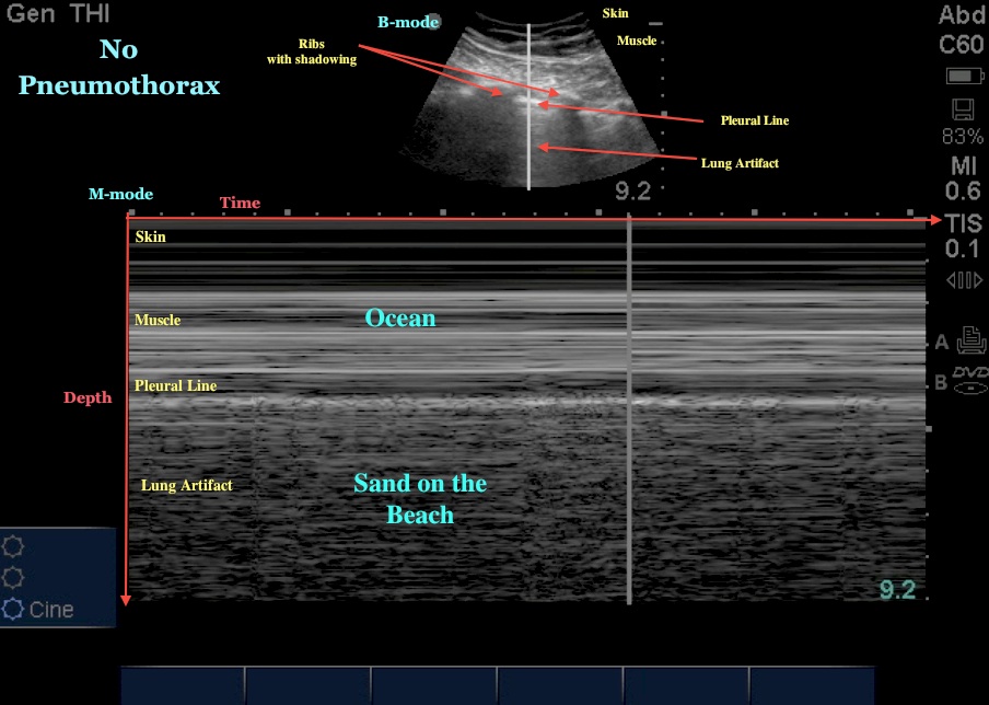
A-mode A-mode (Amplitude-mode) ultrasound is used to judge the depth of an organ, or otherwise assess an organ Modes Ultrasound A-mode- amplitude mode. B-mode- brightness mode. Display of echo amplitude (Y-axis) versus distance (X-axis) into the tissue, which is related to elapsed time and the speed at which ultrasound propagates in the tissue. Sometimes used to calibrate the other modes. Also used to test the symmetry between left and right hemispheres of the brain: R-L then L-R. This form of display (solid areas appear white and fluid areas appear black) is also called gray scale. The B-mode scan is the basis of 2D scanning. The transducer is moved about to view the body from a variety of angles. The probe can be moved in a line (linear scan), or rotated from a particular position (sector scan). System scans frames/s. Hand-held transducer moved to different positions or held at different angles to get complete picture. Transducer can be moved and angles so that get. 3-D information. The simplest type of scanner is just a speeded up version of the 2-D B-scan , allowing a rapid series of still pictures to be built up into a video of the movement. The M-mode (Motion-mode) ultrasound is used for analyzing moving body parts (also called time-motion or TM-mode) commonly in cardiac and fetal cardiac imaging. Used for studying the motion of interface. The high sampling frequency (up to 1000 pulses per second) is useful in assessing rates and motion, particularly in cardiac structures such as the various valves and the chamber walls. Diagnostic Applications: Ultrasound has been used in a variety of clinical settings, including Obstetrics and Gynecology, Endocrinology, Cardiology, Urology, Ophthalmology, Neurology and Musculoskeletal. Endocrinology. In abdominal Sonography, the solid organs of the abdomen are imaged such as the pancreas, aorta, inferior vena cava, liver, gall bladder, bile ducts and spleen.. Determining the position of the fetus to see if it is in the normal head down position. Checking the position of placenta to see if it is improperly developing. Seeing the number of fetuses in uterus. Checking the fetus growth rate by making many measurements. Seeing tumors of breast. Cardiology. To diagnose the dilation of parts of the heart and the function of heart ventricles and valves. Measuring blood flow through the heart and major blood vessels. Measuring the blood flow through the kidney. Seeing the kidney stones. Detecting the prostate cancer. Neurology. For assessing blood flow and stenoses in the carotid arteries (Carotid ultrasonography) and the big intracerebral arteries. Musculoskeletal. Seeing tendons, muscles, nerves, and bone surfaces. Therapeutic applications use ultrasound to bring heat or agitation into the body. Ultrasound may be used to clean teeth in dental hygiene. Ultrasound sources may be used to generate regional heating and mechanical changes in biological tissue, e.g. in physical therapy and cancer treatment. However the use of ultrasound in the treatment of musculoskeletal conditions has fallen out of favor. Focused ultrasound may be used to break up kidney stones by lithotripsy. Ultrasound may be used for cataract treatment by phacoemulsification. Ultrasound scanning is noninvasive (no needles or injections) and is usually painless. Ultrasound is widely available, easy-to-use and less expensive than other imaging methods. Ultrasound imaging uses non ionizing radiation. Ultrasound scanning gives a clear picture of soft tissues that do not show up well on x-ray images. Ultrasound causes no health problems and may be repeated as often as is necessary if medically indicated. There are no hazards for the patient and operator. The major disadvantage is that the resolution of images is often limited. Still in many situations where X-rays produce a much higher resolution. Bone absorbs ultrasound so that brain images are hard to get. Attenuation can reduce the resolution of the image. Sonography performs very poorly when there is a gas between the transducer and the organ of interest. Cont…. Images of tissues on the far side of lungs are impossible to get. Cleaning. This includes the removal of grease, dirt, rust and paint from metal, ceramic, glass and crystal surfaces of parts used in the electronic, automotive, aircraft, and precision instruments industries. Flow Metering. It can be used to monitor closed systems, such as a coolant in a nuclear power plant. Soldering and Welding. Ultrasound has also proved to be very useful for joining plastic materials. It can be used for both soldering and welding. Ultrasound has been used to measure the thickness of fat layers on pigs and cows as part of livestock management. It has also been used in improve the quality of homogenized milk. A related application is pest control, including killing insects. Oceanography. In addition to the tracking of submarines. Oceanographic applications include mapping the contour of the sea bottom, discovering sunken ships. Doppler Ultrasound. Electroencephalograph. Diathermy (Electrosurgery) Lithotripsy (Extracorporeal Shock Wave Lithotripsy) Blood Gas Analyzer. Anesthesia.


.jpg)